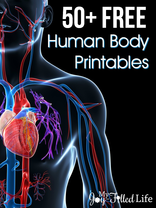
In a world saturated with information, how does one truly see the intricate beauty and complex mechanics of the human form? The answer lies in accessibility: specifically, the abundance of high-quality, freely available human anatomy visuals now at our fingertips.
The digital age has democratized knowledge, and the field of human anatomy is a prime beneficiary. No longer confined to textbooks and expensive medical illustrations, aspiring artists, medical professionals, educators, and curious minds alike now have access to a vast library of anatomical resources. This includes everything from detailed 3D models to meticulously rendered illustrations, all designed to illuminate the inner workings of the human body. The explosion of readily available imagery has revolutionized how we learn, teach, and appreciate the complexities that define us.
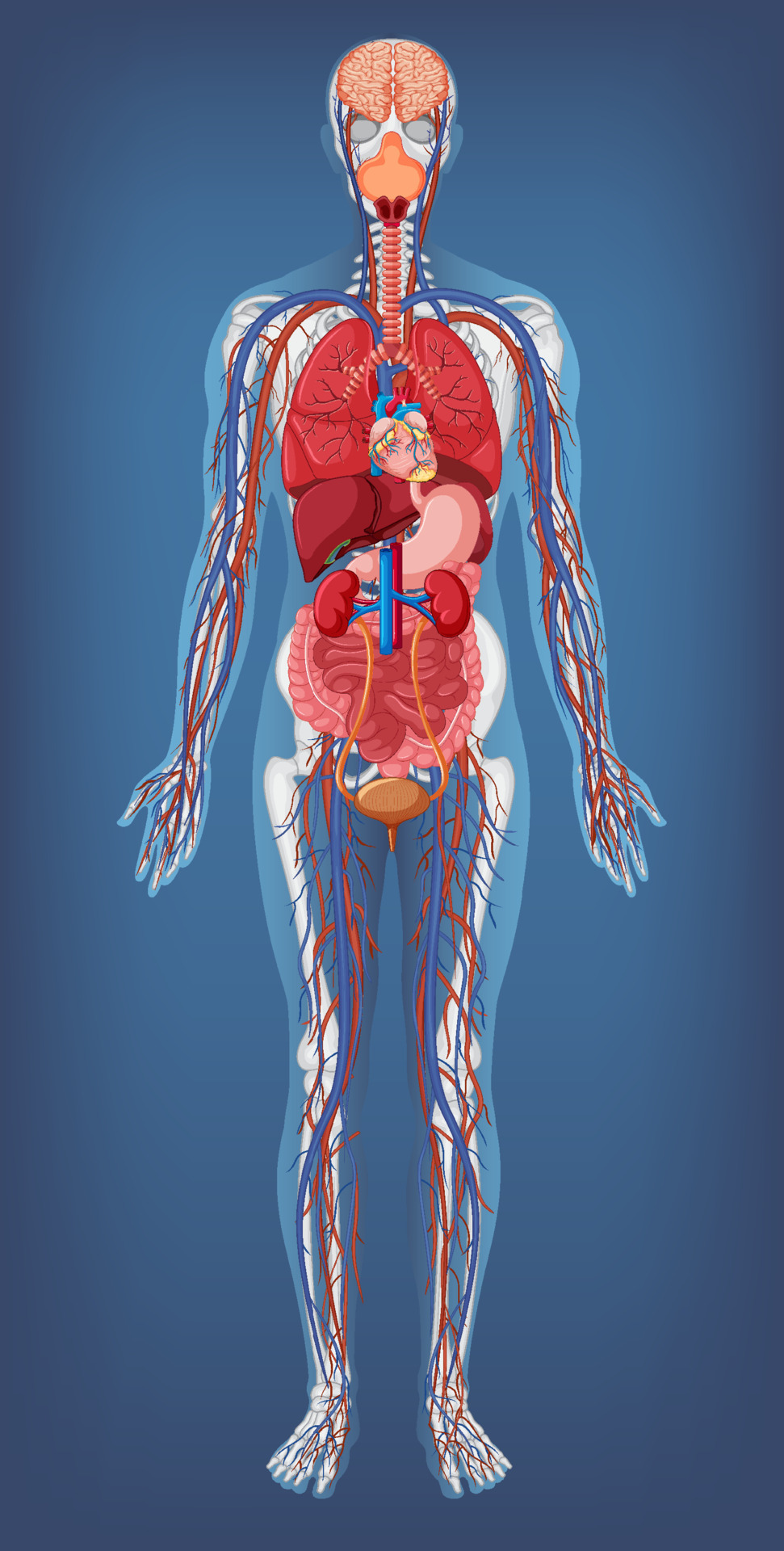
Consider the scale of resources. Thousands upon thousands of free images are at your disposal. A simple search can yield a staggering number of results, from detailed cross-sections and skeletal structures to intricate renderings of the nervous system and the cardiovascular network. Websites offer hundreds of thousands of stock photos, ensuring you can find the perfect image for any project. Medical art libraries are dedicated to providing free, high-quality visuals for medical professionals, students, and anyone interested in learning about human anatomy. This wealth of information, previously locked behind paywalls or accessible only through specialized institutions, is now freely available to anyone with an internet connection.
- X27a1 Rfk Stadium Commanders Return Campus Redevelopment What You Need To Know
- Excel Charts Arrow Charts Easy Guide Tutorials
The benefits of this accessibility are manifold. For medical students, access to these resources can supplement their studies, providing visual aids that clarify complex concepts. Artists, in turn, can use the images as reference, allowing them to create accurate and detailed representations of the human body. Educators can integrate these visuals into their presentations, making lessons more engaging and accessible to students of all levels. Furthermore, the availability of these images promotes a deeper understanding of the human body, fostering a greater appreciation for its intricate design and functions.
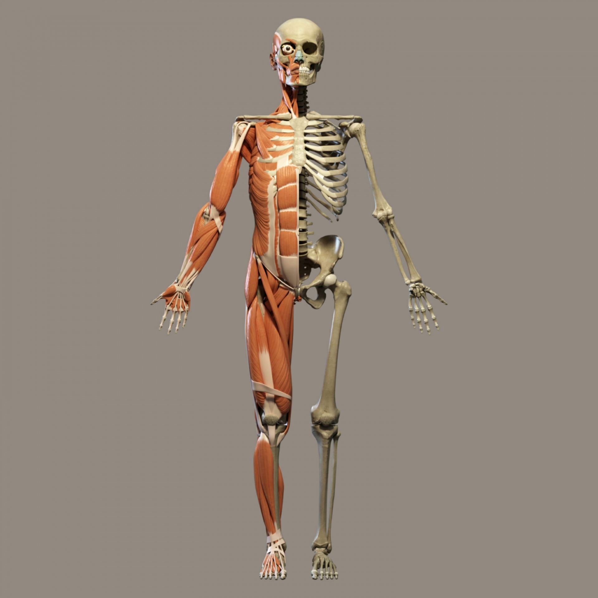
One of the notable advancements is the emergence of 3D models and interactive platforms. Imagine the ability to rotate and dissect a virtual human body, exploring its structures from any angle. Such applications are particularly useful for medical students and practitioners, providing them with an unparalleled opportunity to visualize and understand the complexities of human anatomy. Interactive textbooks, organized by body systems, have also become popular, allowing students to explore the human body in a dynamic and engaging way. This innovative approach to learning transforms anatomy from a series of static diagrams into an immersive and interactive experience.
The ability to download and use these images for free is a critical aspect. Most platforms offer images under Creative Commons licenses, allowing users to copy, modify, distribute, and use the images for commercial purposes without seeking permission or providing attribution. This policy is a testament to the open-source movement and promotes knowledge sharing and creativity. It empowers users to create presentations, publications, videos, quizzes, and flashcards, all enriched by accurate and visually appealing anatomical illustrations.
- Best Single Flower Vases For Any Bloom Decor Shop Now
- Artists Stories John Bramblitt More Art World Insights
The range of available resources extends beyond simple diagrams. Many platforms offer illustrations designed to simplify complex medical concepts, making them ideal for powerpoint presentations, journal publications, and textbooks. Some resources, such as those provided by the National Cancer Institute, offer searchable collections of medical images focused on specific areas of study. The sheer scope of the available imagery ensures that there is something for everyone, from beginners to seasoned professionals.
The practical applications of these resources are vast. Artists can utilize them for reference when drawing the human body, ensuring anatomical accuracy and detail. Medical professionals can incorporate them into patient education materials, allowing them to better explain complex medical conditions. Teachers can create more engaging lessons by incorporating visuals, making difficult topics easier to understand. The availability of free, high-quality images democratizes the ability to study and appreciate the human form.
Consider the meticulous detail of these visuals. Many platforms offer high-resolution images suitable for professional use. These are not low-quality snapshots, but carefully crafted illustrations and 3D models designed to provide accurate representations of anatomical structures. This level of detail is crucial for medical professionals, artists, and anyone who needs to understand the human body in all its complexity.
The availability of these resources is also continually evolving. The data sets are vast, constantly being updated. The male data set, for instance, is approximately 15 gigabytes in size. Higher resolution axial anatomical images continue to be made available, ensuring that the available information is constantly being enhanced. The continuous development ensures the resources remain cutting-edge and relevant.
The scope and scale of accessible resources are astounding. The sheer number of images and illustrations available underscores the importance of readily available anatomical information. The emphasis on creative commons licenses is a testament to a desire to share and to create a more accessible environment for learning. The availability of the imagery makes it easy for anyone to dive into the incredible world of the human anatomy. You can easily navigate, download and utilize the resources to learn about the skeletal, muscular, cardiovascular, digestive, endocrine, nervous, respiratory, immune/lymphatic, urinary, and female reproductive systems.
In this digital age, the democratization of knowledge is reshaping many fields, and none more profoundly than human anatomy. What was once restricted to textbooks and specialized institutions is now accessible to anyone. This transformation offers an unprecedented opportunity to explore, understand, and appreciate the intricacies of the human body. This is the evolution of knowledge. It is available. It is free. And it is waiting to be discovered.
To reiterate the core benefit, there are thousands of free images of human anatomy available. These images are free to download and use. They include a wide range of illustrations and models, designed to support all projects requiring them.
The best resources include access to:
- 25,744 free images of human anatomy.
- 1,944 free images of anatomy.
- 43,214 free images of the human body.
- 600,000+ human anatomy stock photos.
- 11,654+ free human anatomy illustrations.
The process of obtaining these images is often simple. Just click on any image, right-click, and select "save as". Images can also be found through a browse or filter function, allowing you to find the perfect picture for your project.
| Category | Details |
|---|---|
| Resource Type | Free Images & Illustrations of Human Anatomy |
| Availability | Online, Accessible through various websites and databases |
| Licensing | Creative Commons (allowing commercial use, modification, and distribution) |
| Primary Users | Medical professionals, students, artists, educators, and anyone interested in human anatomy |
| Image Formats | Stock photos, illustrations, 3D models, anatomical diagrams, and more. |
| Key Features | High-resolution downloads, accurate anatomical representation, diverse range of subjects and systems, interactive 3D models |
| Purpose | To provide visual aids for learning, teaching, artistic reference, and medical education |
| Examples of use | Presentations, publications, articles, educational materials, artistic renderings, and patient education |
| Main benefits | Free access to high-quality visuals, promotes knowledge-sharing, supports education and medical training, simplifies complex concepts |
| Reference Websites |
|
Detail Author:
- Name : Myrtice Boyle
- Username : kamille.kuhlman
- Email : herman80@bartell.com
- Birthdate : 1972-05-22
- Address : 641 Osinski Inlet Apt. 078 Lake Alfordmouth, PA 71279
- Phone : +1 (586) 929-9619
- Company : Gleason LLC
- Job : Precision Dyer
- Bio : Repudiandae ut at est necessitatibus nihil sunt. Et sunt odit est ipsa maiores. Beatae numquam aperiam enim facere veniam iste facilis expedita.
Socials
linkedin:
- url : https://linkedin.com/in/sokuneva
- username : sokuneva
- bio : Alias adipisci et reprehenderit molestiae.
- followers : 1317
- following : 1120
twitter:
- url : https://twitter.com/sarai2231
- username : sarai2231
- bio : Rem ipsum aliquam velit in optio error est pariatur. Cumque omnis iure architecto et aut sunt. Ab enim atque sequi nostrum. Et voluptas culpa dolor unde earum.
- followers : 6809
- following : 2723
instagram:
- url : https://instagram.com/sokuneva
- username : sokuneva
- bio : Minima labore et ad earum ab quasi. Dignissimos minima nesciunt iure officiis vel.
- followers : 469
- following : 1606
tiktok:
- url : https://tiktok.com/@sarai.okuneva
- username : sarai.okuneva
- bio : Qui tenetur magni praesentium possimus. Rerum aut dolorum provident laboriosam.
- followers : 3316
- following : 357
facebook:
- url : https://facebook.com/sarai_okuneva
- username : sarai_okuneva
- bio : Reiciendis dolor asperiores saepe neque excepturi dignissimos.
- followers : 2191
- following : 110
