Does the intricate dance of the human body's form truly dictate its function, especially in the complex region of the head and neck? Understanding the detailed anatomy of this area is paramount for accurate diagnosis and treatment of a wide range of medical conditions, from vascular lesions to musculoskeletal disorders.
In the realm of medical imaging, the term "head and neck" encompasses all anatomical structures within this region, excluding the central nervous system the brain, spinal cord, and their associated vascular structures and protective membranes. This focused approach is crucial for radiologists and clinicians alike, as it allows for a more precise and efficient evaluation of specific pathologies.
Here's a look at some key facts about head anatomy:
| Structure | Key Features |
|---|---|
| Skull | Protective bony framework for the brain; comprised of the cranium and facial bones. The head rests on the top part of the vertebral column, with the skull joining at C1 (the first cervical vertebra known as the atlas). |
| Cervical Spine | The skeletal section of the head and neck forms the top part of the axial skeleton and is made up of the skull, hyoid bone, auditory ossicles, and cervical spine. Provides support and allows for head movement. |
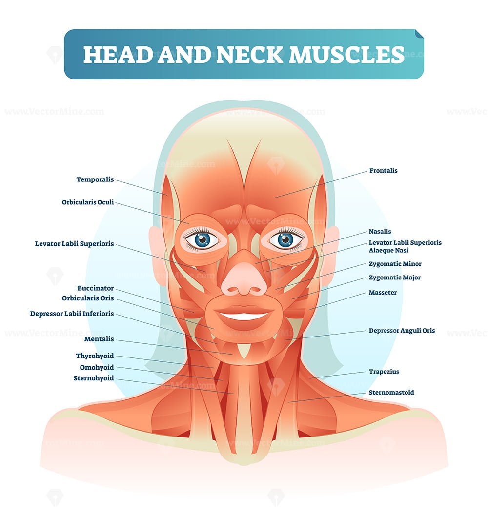
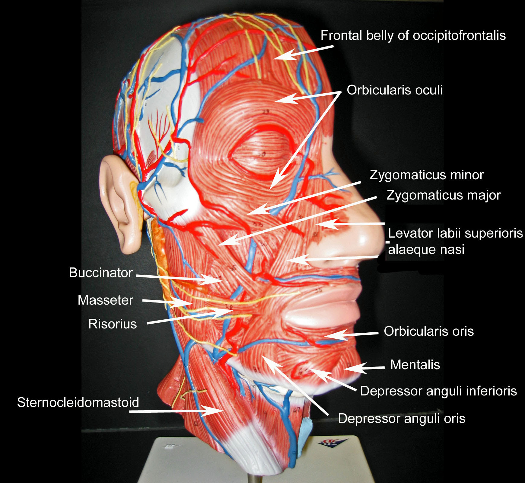
| Muscles of Mastication | Masseter, temporalis, medial pterygoid, and lateral pterygoid muscles are active in grinding actions of chewing. |
| Muscles of the Neck | Sternocleidomastoid, trapezius, and others; responsible for head movement, closely related to major blood vessels, nerves, and elements of the respiratory and gastrointestinal systems. |
| Vascular Structures | Major arteries and veins supplying the head and neck; crucial to investigation of head/neck vascular lesions. |
| Nerves | Cranial nerves, cervical plexus; responsible for sensory input, motor control, and autonomic functions. |
| Lymph Nodes | Facial nodes, cervical nodes, jugulodigastric node, occipital nodes; play a vital role in immune function and can be indicators of disease. |
| Temporomandibular Joint (TMJ) | Capsule and & articular disk of the temporomandibular joint; allows for complex movements of the jaw. |
Specialized imaging techniques like CT angiography are frequently employed to assess for conditions like carotid stenosis, providing detailed visualization of blood vessels and surrounding structures. The ability to interpret these images accurately relies heavily on a strong understanding of the normal anatomical relationships within the head and neck. This knowledge is also essential in understanding where the "foramen" is, a hole in a bone through which blood vessels and nerves pass.
Let's take a deeper dive into some of the specific anatomical regions that are important in the head and neck, and see how they interact:
Bones and Skeletal Structures:
- Yin Yang Koi Fish Meaning Symbolism Tattoo Ideas Explained
- Get Free Blue Soccer Wallpapers Now Download Personalize
The skeletal framework provides the fundamental structure of the head and neck. It's more than just a static support system; it's a dynamic component that facilitates movement, protects vital organs, and serves as attachment points for muscles. Key bony elements to consider include:
- The Skull: Encases and protects the brain. It's composed of the cranium and facial bones, each contributing to the overall structural integrity of the head.
- Cervical Spine: The first seven vertebrae (C1-C7) of the vertebral column that make up the neck, providing support and flexibility. C1, also known as the atlas, articulates with the skull, allowing for a wide range of head movements.
- Hyoid Bone: A unique bone located in the neck that doesn't articulate with any other bone. It serves as an attachment point for muscles involved in swallowing and speech.
- Auditory Ossicles: The small bones in the middle ear (malleus, incus, and stapes) that transmit sound vibrations.
Muscles:
The muscular system is responsible for the movements of the head and neck, allowing for facial expressions, chewing, speech, and head movement. Several muscle groups play crucial roles:
- Muscles of Mastication: Including the masseter, temporalis, medial pterygoid, and lateral pterygoid. They are responsible for chewing.
- Neck Muscles: The sternocleidomastoid and trapezius are particularly important for gross motor movement, pulling the skull and jaw towards the shoulders, spine, and scapula.
- Orbital, Nasal, Oral, Auricular, and Scalp/Neck Muscles: These contribute to a wide array of functions.
Vascular System:
The vascular network of the head and neck is highly complex, providing essential blood supply to all the tissues and organs. Key components include:
- Major Arteries: The carotid arteries and their branches supply oxygenated blood to the head and neck.
- Major Veins: Jugular veins drain deoxygenated blood from the head and neck.
- Dural Venous Sinuses: Important for venous drainage of the brain.
Nervous System:
The nervous system provides the communication network for the head and neck. Key components include:
- Cranial Nerves: Twelve pairs of cranial nerves that originate from the brain and control various functions, including sensation, movement, and autonomic functions.
- Cervical Plexus: A network of nerves that innervates the neck and provides sensory and motor innervation.
Lymphatic System:
The lymphatic system plays a critical role in immune surveillance and fluid balance. Key structures include:
- Lymph Nodes: Found throughout the head and neck, filtering lymph fluid and housing immune cells. Specific locations include facial nodes, cervical nodes, jugulodigastric node, and occipital nodes.
Other Important Structures:
- Temporomandibular Joint (TMJ): Allows for complex jaw movements.
- Upper Respiratory and Digestive Tracts: The pharynx, larynx, and upper esophagus play crucial roles in swallowing, speech, and respiration.
Radiological investigations, such as CT scans and MRIs, are indispensable tools in the diagnosis and management of head and neck conditions. The ability to accurately interpret these images requires a thorough understanding of the normal anatomy and the ability to identify subtle variations or abnormalities.
For example, in the investigation of carotid stenosis, a CT angiogram provides detailed images of the carotid arteries, allowing clinicians to assess the degree of narrowing and plan appropriate treatment. The anatomical knowledge is crucial for the proper interpretation of the CT angiogram images, enabling identification of the "right vertebral artery atlantic (v3) segment" and the "left occipital artery," etc. Another scenario would be a labeled head and neck anatomy study, which would help physicians understand the placement of each part.
The precise location of muscles is also critical in interpreting radiological findings. For example, the masseter, temporalis, medial pterygoid, and lateral pterygoid muscles can be identified on imaging, allowing clinicians to assess their health and any potential abnormalities.
3D interactive models and tutorials, along with detailed charts, further enhance understanding of head and neck anatomy.
Additionally, the lymphatic system within the head and neck can be seen via imaging, which is important to diagnose any disease in that area. Facial nodes, cervical nodes, jugulodigastric nodes, and occipital nodes can all be identified.
The use of detailed charts illustrating normal anatomy and common injuries provides a valuable resource for both clinicians and students. These charts often display multiple views of the head and neck, highlighting the interrelationships between the various systems.
Several resources are available to help study and understand head and neck anatomy. These include textbooks, atlases, online resources, and interactive models. Several people have dedicated their careers to the study of Head and Neck Anatomy:
| Researcher | Specialty | Key Contributions | Website |
|---|---|---|---|
| Arvind Prasad Singh | Radiology | Head and neck radiological anatomy | Arvind Prasad Singh |
| Zeinab Cama | Anatomy | Head and Neck Anatomy | Zeinab Cama |
| Atta ur Rahman | Anatomy | Head and Neck Anatomy | Atta ur Rahman |
| Christian Victor Masangkay | Radiology | CT MR anatomy | Christian Victor Masangkay |
| Ru'a Alahnaf Abuamira | Radiology | Head and neck and vascular anatomy | Ru'a Alahnaf Abuamira |
| Neda Noroozian | Anatomy | Labeled Head and Neck Anatomy | Neda Noroozian |
The anatomical relationships within the head and neck are complex, with various structures interacting and overlapping. For example, the muscles of the neck are closely associated with major blood vessels, nerves, and components of the respiratory and gastrointestinal systems. The cervical plexus, a network of nerves, plays a central role in innervating the neck. It is also important to recognize the terminology of osseous anatomy holes, where a foramen is a hole in a bone through which blood vessels and nerves pass. This becomes particularly important during investigation of head/neck vascular lesions.
The application of this knowledge extends beyond initial diagnosis and treatment. The understanding of anatomy is crucial for surgical planning, allowing surgeons to approach procedures with precision and minimize the risk of complications. Furthermore, a thorough understanding of anatomy is essential for understanding the mechanisms of injury, which informs the assessment of injuries, such as fractures, dislocations, and soft tissue trauma. For instance, in cases of trauma, the detailed anatomy of the head and neck is essential for evaluating the extent of injuries and planning appropriate treatment.
Accurate diagnosis is facilitated by comparing various imaging modalities, such as coronal sections through the orbits, sinuses, and oral cavity, to T1 and T2 MRIs in midsagittal sections. The anatomical images can be overlaid with other views of the head and neck, highlighting how the spine, muscles, tendons, ligaments, and the vascular system function together. The ability to recognize the normal anatomy and understand anatomical variations is also vital for distinguishing between normal and abnormal findings, improving the accuracy of diagnoses and allowing for better patient care.
In conclusion, a comprehensive understanding of head and neck anatomy is critical in the medical field. Whether it's identifying the right vertebral artery or analyzing the muscles, the intricate details of the head and neck are key to providing a safe and informed experience.

Detail Author:
- Name : Harry Lynch
- Username : akoss
- Email : juwan.aufderhar@yahoo.com
- Birthdate : 1986-01-15
- Address : 297 Kaia Manors Suite 015 Lake Elenor, CA 24412
- Phone : +19472605277
- Company : Hills, Mraz and Rosenbaum
- Job : Fraud Investigator
- Bio : Ea quasi laborum vel est aut. Qui praesentium quibusdam autem quae ea labore. Eum dolorem soluta rem laudantium.
Socials
facebook:
- url : https://facebook.com/dewayne_dev
- username : dewayne_dev
- bio : Quia unde repellendus vero ab dolorem adipisci. Magnam id iste ullam ullam ut.
- followers : 1890
- following : 2851
twitter:
- url : https://twitter.com/dewaynebatz
- username : dewaynebatz
- bio : Qui voluptas temporibus et quibusdam voluptas hic quas. Qui est dolorem a non in. Et dolor sit est. Iure harum atque ut.
- followers : 4809
- following : 1941
tiktok:
- url : https://tiktok.com/@dewayne8294
- username : dewayne8294
- bio : Quidem deleniti debitis quos voluptas est.
- followers : 2608
- following : 2757
linkedin:
- url : https://linkedin.com/in/dewayne.batz
- username : dewayne.batz
- bio : Autem unde eum quasi delectus voluptas.
- followers : 6801
- following : 567
instagram:
- url : https://instagram.com/batz1970
- username : batz1970
- bio : Quae quis nihil non cumque culpa. Nostrum doloribus exercitationem occaecati numquam deleniti.
- followers : 5159
- following : 1156

