Can a single image truly tell the story of your lungs, revealing the impact of lifestyle choices and potential health threats? The answer is a resounding yes: a chest X-ray, or a similar form of imaging, acts as a critical window into the very essence of your respiratory health, offering invaluable insights into the condition of your lungs and related structures.
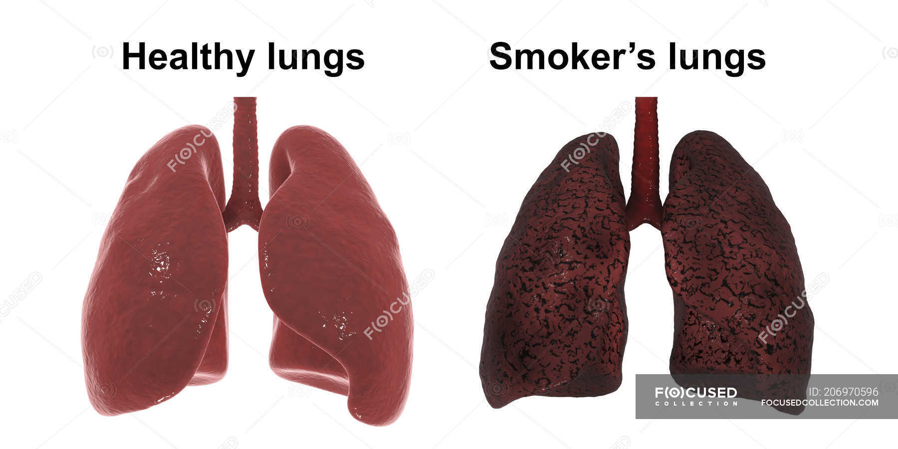
These detailed images are, in essence, windows into the health of your thoracic cavity, providing a glimpse into your lungs, heart, airways, blood vessels, as well as the bones of your spine and chest wall. They are pivotal in distinguishing normal from concerning signs, steering clinicians toward accurate diagnoses and effective treatments. By comparing healthy lungs with those of smokers, one can vividly witness how this habit slowly reduces lung capacity, makes breathing harder, and ultimately causes permanent damage. If you experience any of the symptoms, its crucial to seek medical advice promptly. The radiological assessments provide an essential foundation for understanding a patient's current condition, aiding in early detection and management of various respiratory ailments.
| Aspect | Details |
|---|---|
| Imaging Method: | Chest X-ray |
| Purpose: | To visualize the structures within the chest cavity, including lungs, heart, and major blood vessels. |
| Key Findings: |
|
| Clinical Significance: |
|
| Limitations: |
|
| Reference: | RadiologyInfo.org |
The stark reality is that smoking leaves a lasting effect on your lungs, profoundly changing both their structure and function. This impact is not merely cosmetic; it's a fundamental shift in the very mechanics of breathing and oxygen exchange. The difference becomes glaringly apparent when comparing the healthy lungs on the left with the smoker's lungs, which often appear significantly altered. As one expert observed, "That's a normal looking lung. They're black, they're full of air, just that's what we want to see. That's what your lungs should look like." The contrast is even more pronounced when considering the lungs of a patient battling a serious respiratory illness like COVID-19. These images paint a clear picture of the destructive effects of smoking and severe illnesses.
- Daniel Rohrbough A Columbine Tragedy Remembered Learn More
- X27a1 Rfk Stadium Commanders Return Campus Redevelopment What You Need To Know
Consider the dramatic visual representation. Imagine a side-by-side comparison, reminiscent of a scientific diagram, perhaps titled "2.1," as it might appear in a medical journal. This graphic illustrates the stark differences between healthy lung tissue and lungs exposed to the damaging effects of cigarette smoke. The demo would vividly show a healthy set of lungs beside a set of lungs similar to someone who smoked a pack of cigarettes every day for 20 years. You can literally see the tar that coats the airways in the latter image.
The assessment of lung health using imaging relies on several key principles. First, the comparison of lung zones upper, middle, and lower on both the left and right sides, is crucial. Asymmetry in lung density, represented by either abnormal whiteness (increased density, indicating fluid, or inflammation) or abnormal blackness (decreased density, indicating air trapping or damage), becomes a primary focus.
Lung nodules are another critical area of focus. How they appear on imaging can be a vital diagnostic factor. However, it's important to acknowledge the limitations; these imaging methods may sometimes miss smaller nodules and might fail to fully evaluate the composition of a nodule, requiring further, more detailed investigations. Every radiologist must be an expert in chest film reading, emphasizing the importance of proper interpretation for accurate diagnoses. This skill is paramount in clinical practice, as a misdiagnosis can lead to potentially life-threatening consequences.
- Discover Abandoned Industrial Sites In America Photos Stories
- Unlocking Hip Stability Ligaments Pain Recovery Your Guide
Cigarette smoking is associated with a wide array of abnormalities throughout the body, resulting in both illness and, tragically, all too often, death. Indeed, if all deaths from diseases related to smoking (lung disease, heart disease, and cancers of many different organs) were considered, a compelling argument could be made for cigarette smoking as the leading cause of death in industrialized countries. The case of a patient, let's say in a hospital in a large city, who, despite having no history of smoking, experienced a sudden collapse while waiting for a staging CT scan in radiology, is a stark reminder of the fragility of life and the devastating impact of unexpected medical events. This patient could not be resuscitated, and the gross and microscopic images from the autopsy provided a crucial window into the cause of death.
The importance of seeking medical advice when experiencing symptoms cannot be overstated. A cough that persists, shortness of breath, chest pain, or any other concerning symptom should prompt a consultation with a healthcare professional. Early detection and intervention are critical in managing and treating lung conditions.
This information should not substitute for professional medical advice, diagnosis, or treatment. Always seek the advice of your physician or other qualified health provider with any questions you may have regarding a medical condition. Never disregard professional medical advice or delay in seeking it because of something you have read here.


Detail Author:
- Name : Myrtice Boyle
- Username : kamille.kuhlman
- Email : herman80@bartell.com
- Birthdate : 1972-05-22
- Address : 641 Osinski Inlet Apt. 078 Lake Alfordmouth, PA 71279
- Phone : +1 (586) 929-9619
- Company : Gleason LLC
- Job : Precision Dyer
- Bio : Repudiandae ut at est necessitatibus nihil sunt. Et sunt odit est ipsa maiores. Beatae numquam aperiam enim facere veniam iste facilis expedita.
Socials
linkedin:
- url : https://linkedin.com/in/sokuneva
- username : sokuneva
- bio : Alias adipisci et reprehenderit molestiae.
- followers : 1317
- following : 1120
twitter:
- url : https://twitter.com/sarai2231
- username : sarai2231
- bio : Rem ipsum aliquam velit in optio error est pariatur. Cumque omnis iure architecto et aut sunt. Ab enim atque sequi nostrum. Et voluptas culpa dolor unde earum.
- followers : 6809
- following : 2723
instagram:
- url : https://instagram.com/sokuneva
- username : sokuneva
- bio : Minima labore et ad earum ab quasi. Dignissimos minima nesciunt iure officiis vel.
- followers : 469
- following : 1606
tiktok:
- url : https://tiktok.com/@sarai.okuneva
- username : sarai.okuneva
- bio : Qui tenetur magni praesentium possimus. Rerum aut dolorum provident laboriosam.
- followers : 3316
- following : 357
facebook:
- url : https://facebook.com/sarai_okuneva
- username : sarai_okuneva
- bio : Reiciendis dolor asperiores saepe neque excepturi dignissimos.
- followers : 2191
- following : 110
