Is abdominal pain a mystery you're trying to solve? An abdominal ultrasound can offer a quick and non-invasive glimpse into the inner workings of your abdomen, potentially revealing the source of your discomfort and providing crucial information for your doctor to make an accurate diagnosis.
The world of medical imaging offers a variety of tools to peer inside the human body, and among these, the abdominal ultrasound stands out for its accessibility and safety. This diagnostic technique utilizes sound waves, a form of energy that poses no risk of radiation exposure, making it a preferred choice for many patients, including pregnant women and children. Unlike X-rays or CT scans, which employ ionizing radiation, an abdominal ultrasound relies on the principles of echolocation, similar to how bats navigate in the dark. High-frequency sound waves are emitted from a handheld device called a transducer, which is gently moved over the skin of the abdomen. These sound waves travel through the tissues and organs, bouncing back in the form of echoes. The transducer captures these echoes, and a computer processes them to create real-time images that display the internal structures of the abdomen on a screen.
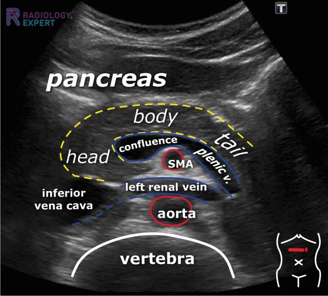
An abdominal ultrasound is a versatile tool, capable of providing detailed images of a wide array of abdominal organs and structures. Among the areas that can be examined are the liver, gallbladder, pancreas, spleen, kidneys, adrenal glands, and the abdominal aorta, the major blood vessel that carries blood to the lower part of the body. The exam typically takes around 30 minutes to complete, offering a relatively quick and efficient way to assess the health of these vital organs.
- Aaron Brooks Stats Career Raiders Highlights What You Need To Know
- Naked Muscle Men Hot Videos Pics Explore Now
The information gathered from an abdominal ultrasound can be crucial in diagnosing and monitoring a variety of medical conditions. It can help identify gallstones, detect abnormalities in the liver, such as tumors or inflammation, and assess the size and shape of the kidneys and other organs. Furthermore, it can be used to evaluate blood flow through the abdominal aorta, as well as identify potential issues within the abdominal wall.
The process of an abdominal ultrasound is generally straightforward. A specially trained technologist performs the procedure. The patient lies on an examination table, and a gel is applied to the skin of the abdomen. This gel helps to transmit the sound waves from the transducer into the body and to create clearer images. The technologist then moves the transducer over the abdomen, capturing images from various angles. During the exam, the technologist may ask the patient to hold their breath or change positions to get a better view of the organs.
Once the exam is complete, the technologist may ask the patient to dress and wait while they review the ultrasound images. The images and a detailed report are then sent to a radiologist or another specialist for interpretation. The report will include images from the scan, as well as the assessment made by the radiologist, ultrasound technician or other specialist. The report may contain technical medical terminology and other complex data.
- Top Music Producer Wallpapers Download Free Hd Backgrounds Now
- Best Mexican Haircuts Hairstyles For Men Trendy Looks
In some cases, your doctor may order a Doppler ultrasound in conjunction with the abdominal ultrasound. A Doppler ultrasound makes images of blood flow through vessels that run through the abdomen, such as the aorta.
One of the advantages of abdominal ultrasounds is that you should be able to return to your regular activities right after the exam. The procedure is non-invasive, and there are no lingering effects.
The use of abdominal ultrasound is not limited to adults. It is a safe and effective method for evaluating the health of children as well. In pediatric cases, abdominal ultrasounds can be used to diagnose a variety of conditions, such as appendicitis, kidney problems, and liver abnormalities.
Ultrasound technology has advanced significantly over the years. Modern ultrasound machines offer high-resolution imaging and a variety of features that enhance diagnostic accuracy. They can produce detailed images, allowing doctors to see subtle changes in organs and tissues.
Unlike some other imaging tests, ultrasounds don't use ionizing radiation. Instead, they use sound waves to create digital images of the abdominal organs, which can then be stored and shared with other healthcare providers. This makes them a safer option for pregnant women and children.
The ultrasound report includes images from your scan, as well as the assessment made by the radiologist, ultrasound technician or other specialist. The report may contain technical medical terminology and other complex data.
The technologist performing the ultrasound plays a critical role in the quality of the images. An ultrasound is more dependent on the skill of the technologist than other tests.
The information gathered from an abdominal ultrasound can be crucial in diagnosing and monitoring a variety of medical conditions. It can help identify gallstones, detect abnormalities in the liver, such as tumors or inflammation, and assess the size and shape of the kidneys and other organs. Furthermore, it can be used to evaluate blood flow through the abdominal aorta, as well as identify potential issues within the abdominal wall.
Abdominal wall hernias, which can be divided into primary hernias and incisional hernias, are another area that ultrasound can help diagnose. Primary hernias are hernias located at certain weak spots in the abdominal wall, such as in the midline (umbilical and epigastric hernias) and laterally (spigelian and lumbar hernias).
An abdominal ultrasound is just one method used to test for many diseases and conditions.
When screening for HCC (Hepatocellular Carcinoma) is requested, additional steps may be added to the exam, such as linear images of the capsule of the liver to show the degree of nodularity.
When these sound waves are aimed at your body from a small, handheld device called a transducer, these echoes are then translated by a computer into a visual.
In the realm of obstetrics, abdominal ultrasounds have a significant role. An abdominal ultrasound can usually detect a baby's heartbeat if you are at least 8 weeks into your pregnancy.
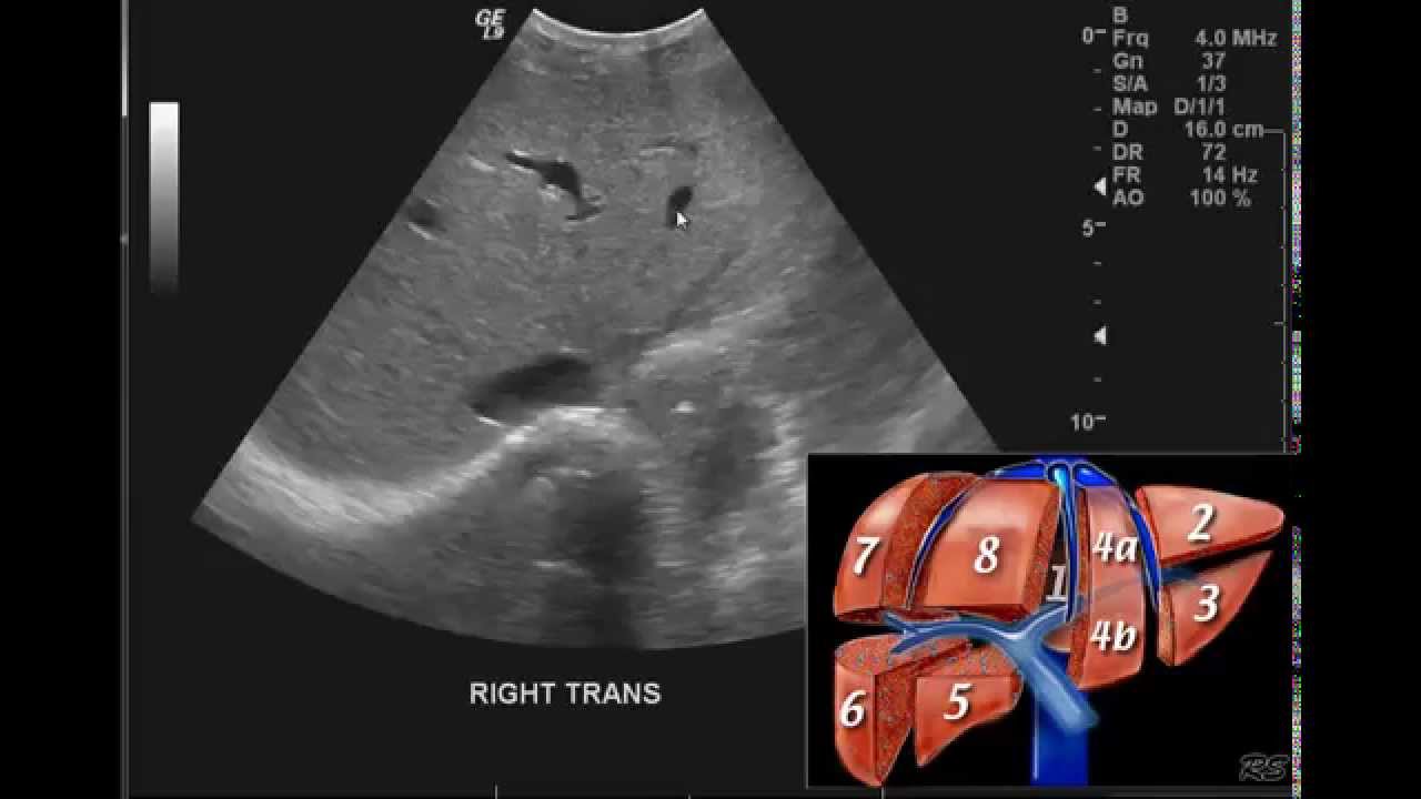
The normal anatomy of the liver appears as a homogeneous, hypoechoic or isoechoic structure on an ultrasound.
Areas that can be checked during an abdominal ultrasound include the: the liver, gallbladder, pancreas, spleen, adrenal glands, kidneys, and abdominal aorta.
A doppler ultrasound is often done alongside an abdominal ultrasound to make images of blood flow through vessels that run through the abdomen, such as the aorta. This large blood vessel passes down the back of the chest and belly and supplies blood to the lower part of the body.
Adding images to a complete order still charges as abd complete. For example, if the exam shows hydronephrosis or pelvicaliectasis (more than a prominent renal pelvis), additional images should be added, such as a representative bladder image (without needing to do volume), and showing the jet only if readily seen; also if the bladder is full, see if dilatation persists after void.
Ultrasound provides a unique role in evaluating internal anatomy.
When it comes to screening for HCC, the following should be added to the exam: linear images of the capsule of the liver to show the degree of nodularity.
Outpatient protocols typically involve performing a full protocol regardless of recent prior imaging.
An abdominal ultrasound exam typically takes about 30 minutes to complete, offering a relatively quick and efficient way to assess the health of these vital organs.
An abdominal ultrasound is done by a specially trained technologist. It does not use radiation but instead uses sound waves which enter the body and then are reflected back by tissues and produce an image on the ultrasound machine.
If your pregnancy has a gestational age of less than 8 weeks (between 6 and 8 weeks), an abdominal ultrasound might be able to detect the babys heartbeat.
An abdominal ultrasound takes pictures of the organs and other structures in your upper belly. It uses sound waves to show images on a screen.

Detail Author:
- Name : Harry Lynch
- Username : akoss
- Email : juwan.aufderhar@yahoo.com
- Birthdate : 1986-01-15
- Address : 297 Kaia Manors Suite 015 Lake Elenor, CA 24412
- Phone : +19472605277
- Company : Hills, Mraz and Rosenbaum
- Job : Fraud Investigator
- Bio : Ea quasi laborum vel est aut. Qui praesentium quibusdam autem quae ea labore. Eum dolorem soluta rem laudantium.
Socials
facebook:
- url : https://facebook.com/dewayne_dev
- username : dewayne_dev
- bio : Quia unde repellendus vero ab dolorem adipisci. Magnam id iste ullam ullam ut.
- followers : 1890
- following : 2851
twitter:
- url : https://twitter.com/dewaynebatz
- username : dewaynebatz
- bio : Qui voluptas temporibus et quibusdam voluptas hic quas. Qui est dolorem a non in. Et dolor sit est. Iure harum atque ut.
- followers : 4809
- following : 1941
tiktok:
- url : https://tiktok.com/@dewayne8294
- username : dewayne8294
- bio : Quidem deleniti debitis quos voluptas est.
- followers : 2608
- following : 2757
linkedin:
- url : https://linkedin.com/in/dewayne.batz
- username : dewayne.batz
- bio : Autem unde eum quasi delectus voluptas.
- followers : 6801
- following : 567
instagram:
- url : https://instagram.com/batz1970
- username : batz1970
- bio : Quae quis nihil non cumque culpa. Nostrum doloribus exercitationem occaecati numquam deleniti.
- followers : 5159
- following : 1156
